 26/07/24
26/07/24
26/07/24
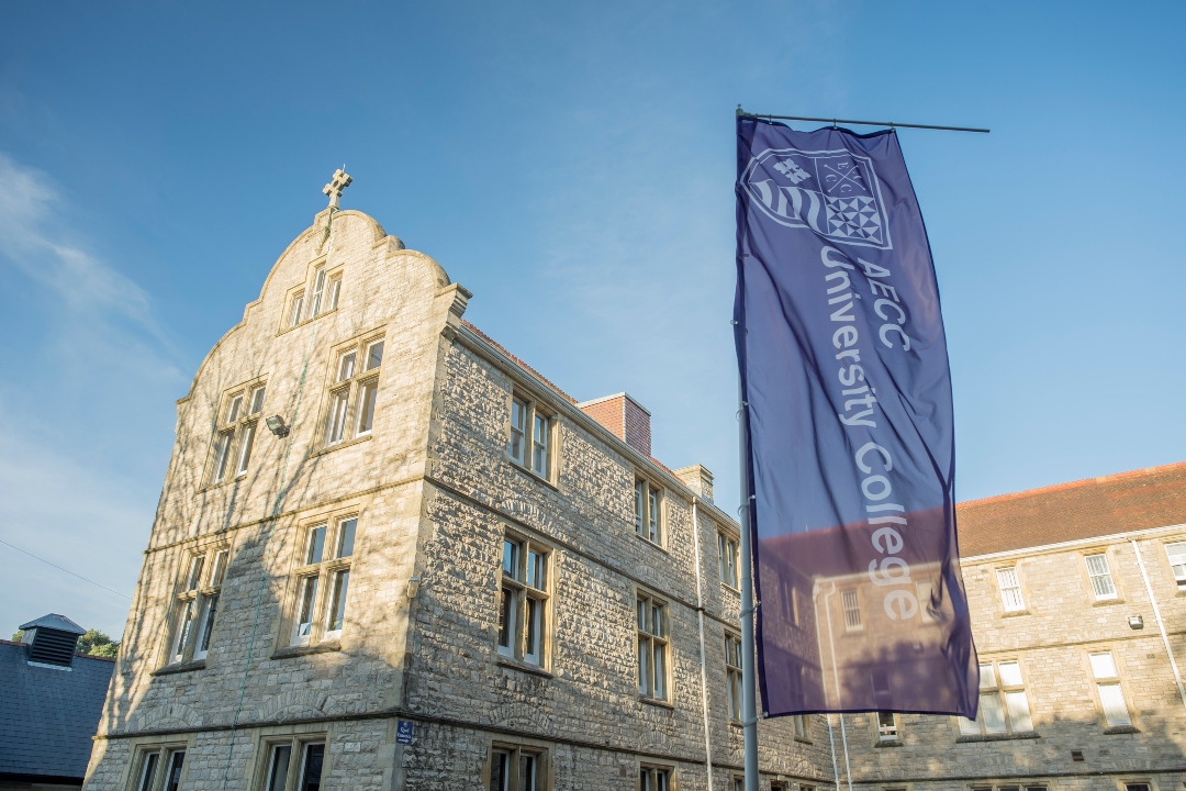
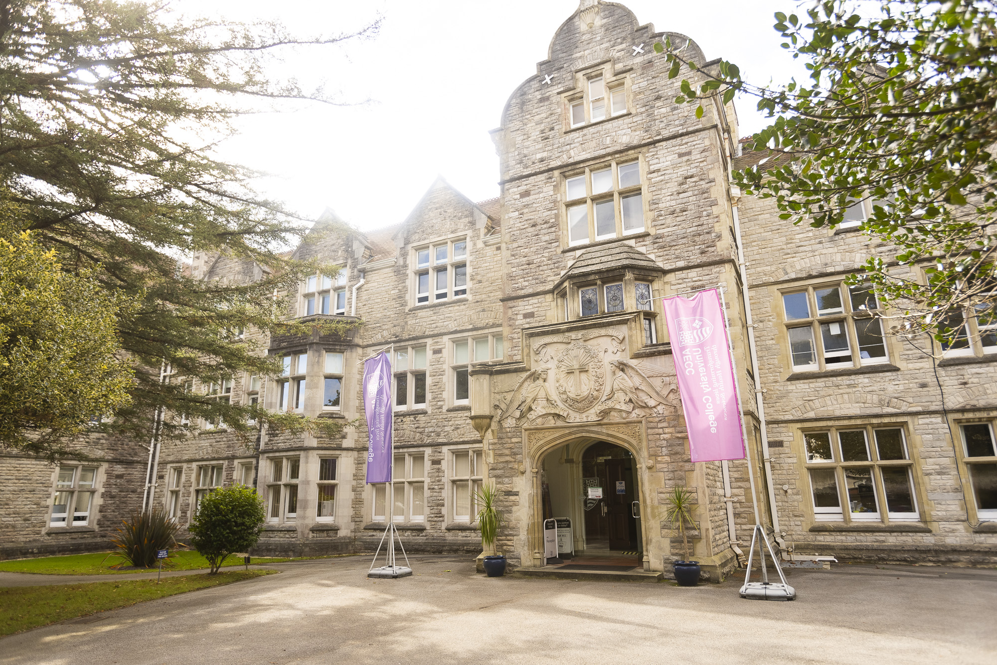
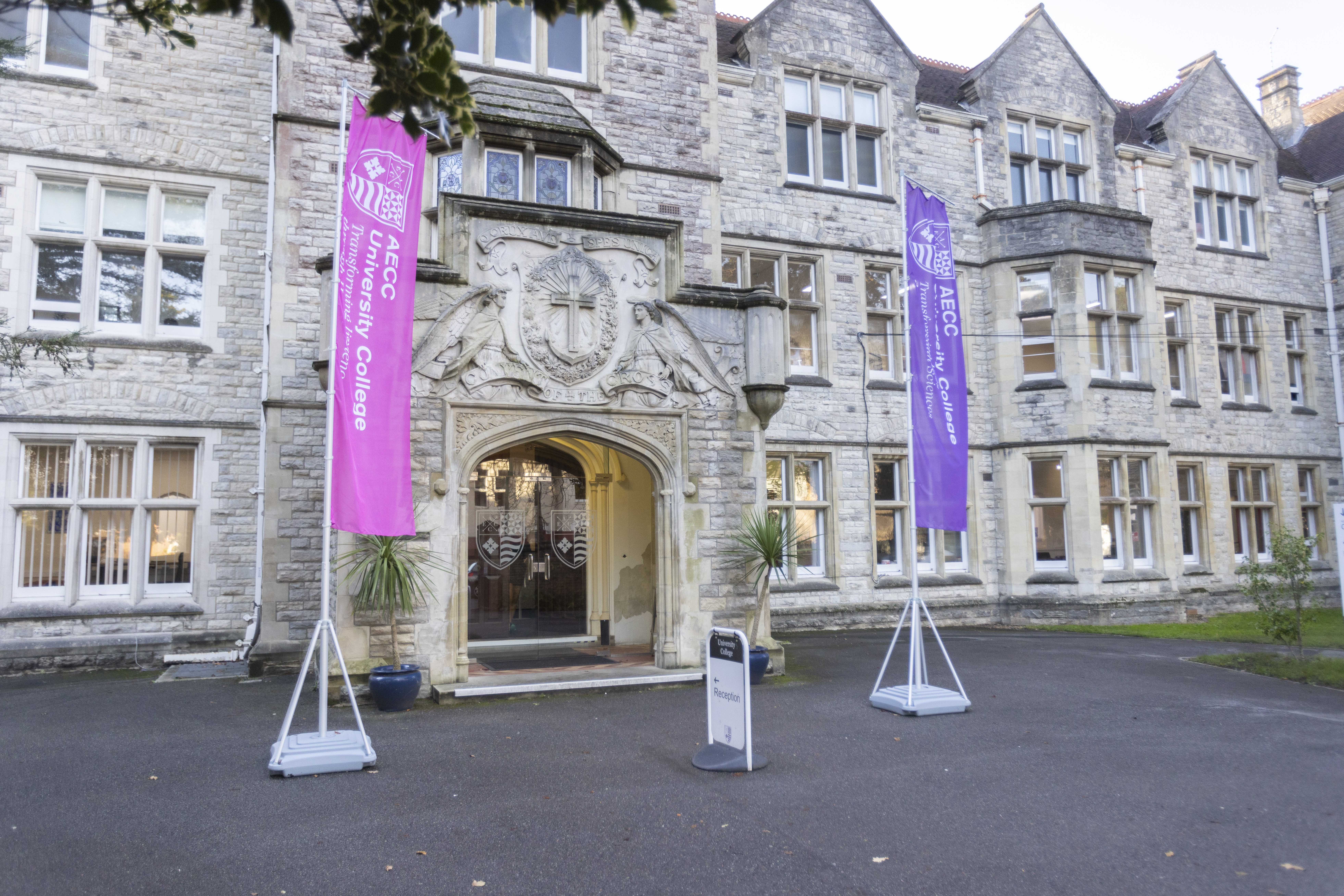
AECC University College
Parkwood Campus
Parkwood Road
Bournemouth
BH5 2DF